news
26 Sept - 1 Oct 2022
the FMU hosts the FASE Basic Course and Symposium for the European Forensic Anthropology Society
28 September 2019
the Unit of forensics organizes th 2nd Forensic Medicine Conference
the FMU hosts the FASE Basic Course and Symposium for the European Forensic Anthropology Society
28 September 2019
the Unit of forensics organizes th 2nd Forensic Medicine Conference
Research / Projects
Virtopsy.GR: Applying imaging methods on death investigation
The purpose of Forensic Science is to record, analyze and interpret scientific medical findings on sudden and violent deaths and to present them clearly to the Court. However, the description of the findings is subjective and dependent on the observer. Imaging techniques have been shown to have remarkable development in recent years, with proven valuable applications in forensic investigation of death ranging from methods of diagnosis, identification to the reconstruction of death conditions. In the context of the general acceptance of imaging techniques in forensic practice, the present study proposes studying forensic cases by using computed tomography and comparing the findings with the results of classical autopsy.

Principal Investigator: Elena Kranioti, Assistant Professor of Forensic Medicine
Cretan Brain Bank (CBB)
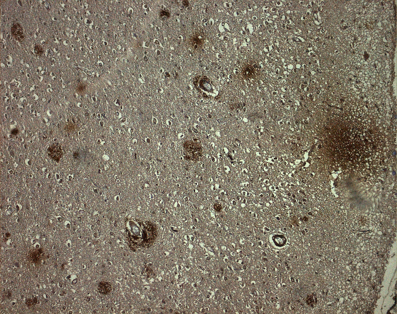
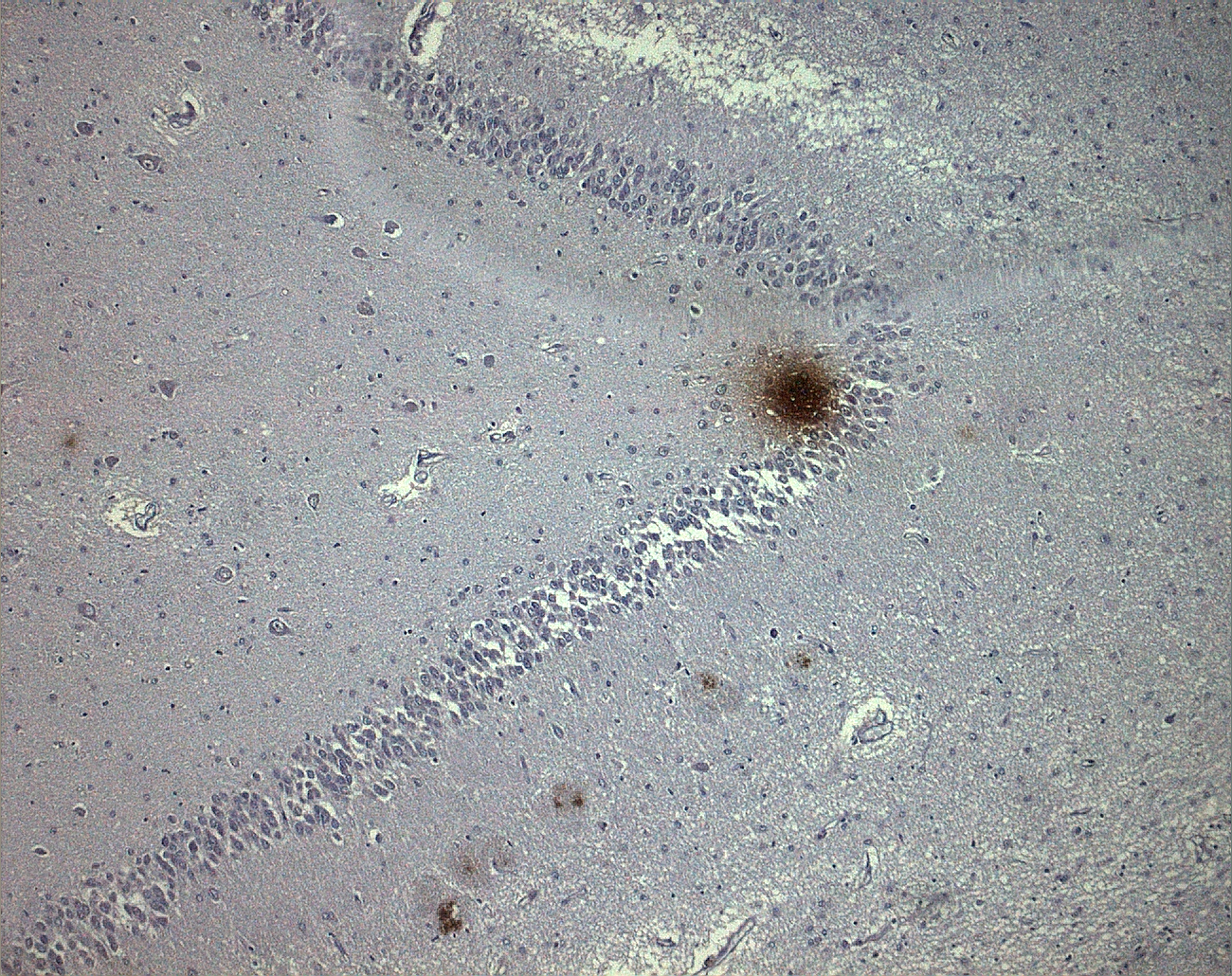
Research in human brain diseases and especially in Alzheimer’s disease (AD), is usually carried out on cell cultures and animal models. However, despite their significant contribution to our understanding of AD, these are not sufficient to fully elucidate its pathophysiology. The study of postmortem brain tissue from patients suffering from AD is crucial for understanding disease mechanisms. In this study, we describe the establishment of a brain bank (Cretan Brain Bank; CBB) focused on AD in Crete, Greece. CBB builds on an ongoing collaboration between the University of Crete, Medical School, and the AD Association of Heraklion (“Alilegii”). Our aim is to advance research on human neurodegeneration using the CBB as a tool. The study was approved by the Ethics Committee of the University Hospital of Heraklion and all subjects and their legal representatives give informed consent.


Principal Investigator: John Zaganas, Assistant Professor of Neyrology
Stable Isotopes and Forensic Applications: Origin of Missing Immigrants
More
-- under construction--
Experimental forensics
-- under construction--


